Oral batyl alcohol supplementation rescues decreased cardiac conduction in ether phospholipid-deficient mice.
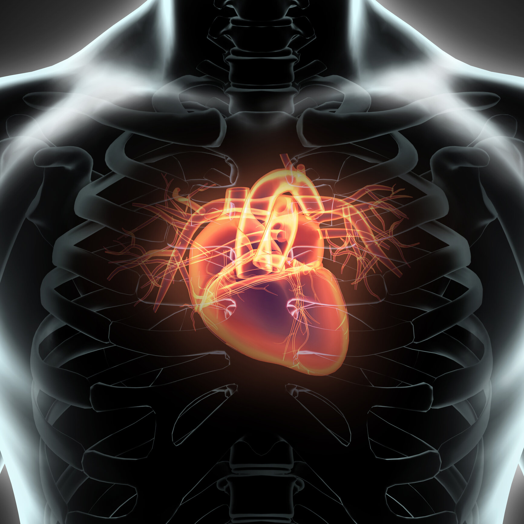
Photo by yodiyim/iStock / Getty Images
Todt H, Dorninger F, Rothauer PJ, Fischer CM, Schranz M, Bruegger B, Lüchtenborg C, Ebner J, Hilber K, Koenig X, Erdem FA, Gawali VS, and Berger J. (2020). Oral batyl alcohol supplementation rescues decreased cardiac conduction in ether phospholipid-deficient mice. J. Inherit. Metab. Dis.
Plasmalogens are a class of phospholipids containing a vinyl-ether bond at the sn-1 position on a glycerol backbone. This double bond gives plasmalogens antioxidant properties and a compact conformation, giving them a unique role in membrane structure and function. Rhizomelic chondrodysplasia punctata (RCDP) is a rare disease caused by mutations in the proteins involved in plasmalogen synthesis. RCDP presents as severe dwarfism, cognitive defects, cataracts, and respiratory complications. Based on two small clinical studies it appears that between 67-75% of patients are diagnosed with some form of congenital heart disease (1, 2), although this is not often recognized as a common characteristic of RCDP. While these studies would typically be viewed as an insufficient sample size (14 & 18 patients per study) to state conclusions from, the prevalence of RCDP is 1:100,000 live births, with less than 100 known cases in North America, therefore this represents a substantial proportion of patients. The effects of plasmalogen deficiency in heart defects found in RCDP is possibly due to the role of plasmalogens in membrane fluidity and lipid rafts, as well as their large contribution to the cell membrane in striated muscle cells. Previous studies using embryonic fibroblasts from the glyceronephosphate o-transferase (Gnpat) knockout (KO) mouse model, which lacks the first enzyme in the plasmalogen biosynthetic pathway, also showed a reduction in connexin 43 (Cx43) protein levels. Cx43 is the most broadly expressed transmembrane protein involved in the formation of gap junctions, which are specialized intercellular connections that directly connect the cytoplasm of two cells and are essential in cardiac ventricles for proper conduction of electrical pulses. Todt et al expanded on these findings using 14 wild-type and 14 Gnpat KO mice to analyze Cx43 expression, ventricular conduction through electrocardiogram (ECG) recordings, and the effects of 2% (w/w) 1-O-rac-octadecylglycerol (batyl alcohol, BA), a plasmalogen precursor, on plasmalogen level and ECG recording.
To determine whether there was any difference in Cx43 expression in Gnpat KO and wild-type mice due to plasmalogen deficiency, heart tissue from embryonic (day 18.5) and aged mice (14-16.5-month-old) were compared using Western blots. No difference could be seen between Cx43 expression in embryonic heart tissue, but in the aged Gnpat KO mice there was a ~40% reduction in Cx43. In addition, to determine if there were any differences in ventricular conduction between the two genotypes, ECG recordings were measured from the four extremities averaged over 100 beats and the longest QRS interval (combination of three graphical points to show ventricular depolarization) was compared. The QRS interval was prolonged in the Gnpat KO mice, increasing the duration of the interval by 13.7% when compared to the wild-type measurements. This extension indicates that the Gnpat KO cardiac conduction is not as efficient as the wild-type heart.
Todt et al analyzed whether treatment with BA for two months could alter the cardiac phenotype of the Gnpat KO mouse. BA treatment was found to rescue ethanolamine plasmalogen (PlsEtn) levels in the Gnpat KO heart tissue to that seen in the wild-type hearts, indicating that the plasmalogen precursor was able to be incorporated into these tissues. As well, the QRS duration was significantly reduced in the Gnpat KO mice, reaching levels seen in the wild-type mice, but no change was observed for the wild-type mice between the untreated and the BA-treated groups.
In addition to the many well-established roles of plasmalogens, this novel work by Todt et al demonstrates the association of plasmalogen deficiency with cardiac conduction and that this defect can benefit from plasmalogen augmentation. Intraventricular conduction delay, seen through the increased duration of QRS in the ECG, is associated with heightened mortality in people with a variety of heart conditions and within the general population. However, it is important to note that the cardiac defects previously described in RCDP patients are developmental in nature, producing anatomical differences in the organ itself, where this article has described a cellular alteration affecting cardiac transduction in the Gnpat KO mouse model. At the time that this blog is published, the molecular cause behind the benefit of the BA treatment is unknown, although has been hypothesized by Todt et al to occur from normalizing the structure of the cardiac cell membranes. Another possible explanation for the role of plasmalogens in cardiac function is their involvement in signal transduction, a similar process to neurotransmission, which is known to be impacted by plasmalogens content, as both are responsible for transmitting signals between cells. Further research could provide more answers on the role of plasmalogens in signal transduction of these cardiac cells, because although this change does appear to be recovered in the BA treated Gnpat KO mice, other differences in the ECG recordings still appear to present between this treated group and the wild-type mice (see figure below). This work provides an expanded role of plasmalogens in transmitting signals and a functional outcome to analyze, however it cannot be directly compared to the human phenotype produced by plasmalogen deficiency. Another important note is that the mice in this study were treated in excess of 2500 mg/kg, depending on how much food was consumed, which is a substantial plasmalogen dose to receive chronically in the case of RCDP patients. Future work looking at the effect of plasmalogen precursors such as BA or complete plasmalogens would be necessary to determine the specific role that plasmalogens have in cardiac function.
1) Huffnagel, IC, Clur SB, Bams-Mengerink AM, Blom NA, Wanders RJA, Waterham HR, and Poll-The BT. (2013) Rhizomelic chondrodysplasia punctata and cardiac pathology. J. Med.Genet., 50: 419-424.
2) Duker AL, Eldridge G, Braverman NE, and Bober MB. (2015) Congenital heart defects common in Rhizomelic Chondrodysplasia Punctata. Am J Med Genet, Part A 170A: 270–272.

Original ECG recording from a WT (blue; upper trace) and a Gnpat KO mouse before treatment with BA (red, middle trace) and after treatment with BA (green, lower trace). The traces (lead I) are aligned with respect to the start of the QRS complex. With reference to the trace in the upper panel (blue), the solid vertical lines indicate, from left to right, the beginning of the P-wave and the beginning and the end of the QRS complex. Furthermore, the peak of the first T-wave (dotted line) and the end of the second T- wave (right) in the upper trace (WT) are indicated. The arrows indicate the end of the QRS complex in the middle trace (Gnpat KO at baseline, red) and the lower trace (Gnpat KO after BA, green). Clearly, the duration of the QRS interval and the QT1 interval are prolonged in the Gnpat KO mouse. However, treatment with BA in this mouse results in normalization of the QRS duration.